Warthin's Tumor : Differential Diagnosis


Comments:
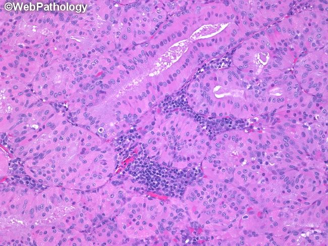
The diagnosis of Warthin’s tumor (WT) is straight-forward in most cases. Cases with a predominant oncocytic cell component may raise suspicion for oncocytoma, mucoepidermoid carcinoma, oncocytosis or acinic cell carcinoma. When squamous and mucinous cell metaplasia is present in WT, its distinction from mucoepidermoid carcinoma may be challenging. Mucoepidermoid carcinomas often carry translocations t(11;19) and t(11;15) resulting in CRTC1-MAML2 and CRTC3-MAML2 fusion genes – a finding not seen in Warthin’s tumor. This image shows classic appearance of Warthin’s tumor with oncocytic epithelium arranged in tubules and nests with associated lymphoid infiltrate.


