
Warthin's Tumor : Fine Needle Aspiration
Home
 Head & Neck
Salivary Glands
Benign Neoplasms of Salivary Glands - I
Warthin's Tumor : Fine Needle Aspiration
Head & Neck
Salivary Glands
Benign Neoplasms of Salivary Glands - I
Warthin's Tumor : Fine Needle Aspiration

slide 72 of 110


Comments:
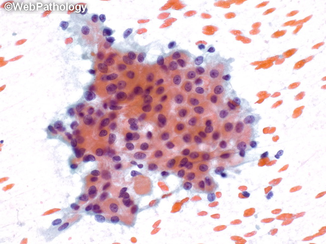
Fine needle aspirate (FNA) smear of a Warthin�s tumor (WT) showing cohesive sheets and clusters of oncocytic cells arranged in a honeycomb pattern. The background usually shows scattered lymphocytes, although they are not seen in this image (PAP stain). Prior manipulation or FNA can cause secondary changes in WT. They include cytologic atypia, metaplasia (squamous and mucous), hemorrhage, infarction, necrosis, acute and chronic inflammation, fibrosis, granulation tissue formation and changes of pseudoinvasion.
slide 72 of 110