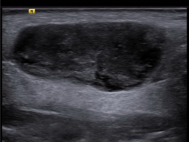
Warthin's Tumor


Comments:
Warthin’s tumor involving the left parotid in a 70 y/o male. Within the left parotid gland there is a large 3.0 x 1.5 x 1.3 cm well- defined hypoechoic lesion (shown in this image) with a smaller adjacent similar lesion. The larger lesion demonstrates mild heterogeneity and internal vascularity (see next image). A core biopsy of this lesion showed features diagnostic of Warthin's tumor. Case courtesy of A.Prof Frank Gaillard, Radiopaedia.org. From the case rID: 26057


