
Pneumocystis Pneumonia : Microscopic Features
Home
 Infectious Disease
Yeast & Yeast-like Fungi
Pneumocystosis
Pneumocystis Pneumonia : Microscopic Features
Infectious Disease
Yeast & Yeast-like Fungi
Pneumocystosis
Pneumocystis Pneumonia : Microscopic Features

slide 6 of 12


Comments:
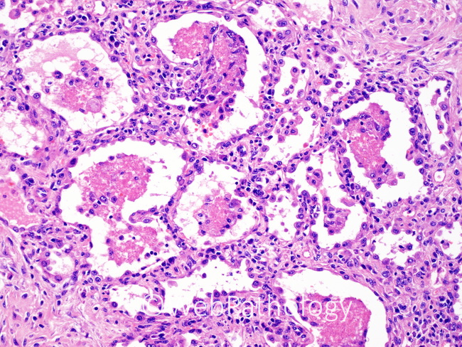
This image shows the classic features of pneumocystis pneumonia - foamy alveolar casts, type 2 pneumocyte hyperplasia, and moderate lymphoplasmacytic interstitial infiltrate. However, more recently, a number of atypical or unusual patterns have been recognized, including acute diffuse alveolar damage with hyaline membranes (see image 8), minimal or no foamy exudates, organizing phase with paucity of organisms, and necrotizing or non-necrotizing granulomatous inflammation that can mimic mycobacterial or other fungal infections. Some cases show cavitary disease, solid fibrotic nodules, and dystrophic calcifications.
slide 6 of 12