
Pneumocystis Pneumonia : Differential Diagnosis


Comments:
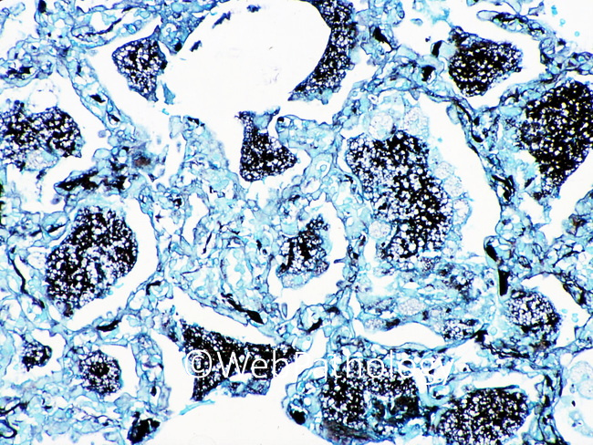
The differential diagnosis of pneumocystis jirovecii is mainly with other small yeast and yeast-like fungi (candida, histoplasma, cryptococcus, blastomyces, coccidioides). Granulomatous pneumocystis is likely to be mistaken for histoplasma, as this feature is expected in histoplasmosis and unexpected in pneumocystosis. Features favoring pneumocystis include the presence of intracystic bodies and the absence of budding. The endospores of Coccidioides can mimic pneumocystis. The presence of immature and mature spherules and the absence of intracystic bodies favor coccidioides. This GMS stained section of lung from a renal transplant recipient shows dense clusters of Pneumocystis jiroveci in alveolar casts. Besides GMS stain, the organisms can be detected by staining with calcofluor white, direct fluorescent antibody test, and real-time PCR.