
Pneumocystis Pneumonia : GMS Stain


Comments:
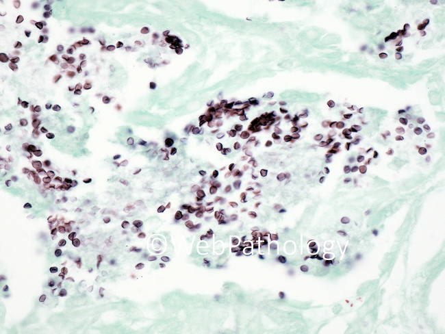
Pneumocystis Pneumonia - GMS Stain: The three stages in the life cycle of pneumocystis are sporozoites, trophozoites, and cysts. The most commonly recognized stage is the cyst. With GMS stain, cysts are 4-7 μm in size, non-budding with oval, crescentic, collapsed, or helmet-like shapes. They have also been likened to crushed ping-pong balls. They are seen in moderate to numerous numbers within foamy alveolar casts. The presence of centrally-located intracystic dots or paired-comma structures helps distinguish pneumocystis jiroveci from other yeast-like organisms (histoplasma, acapsular cryptococcus, candida) and red blood cells. The sporozoites and trophozoites are more readily appreciated in touch imprints and in cytologic preparations of respiratory samples.