
Pneumocystis Pneumonia : Introduction


Comments:
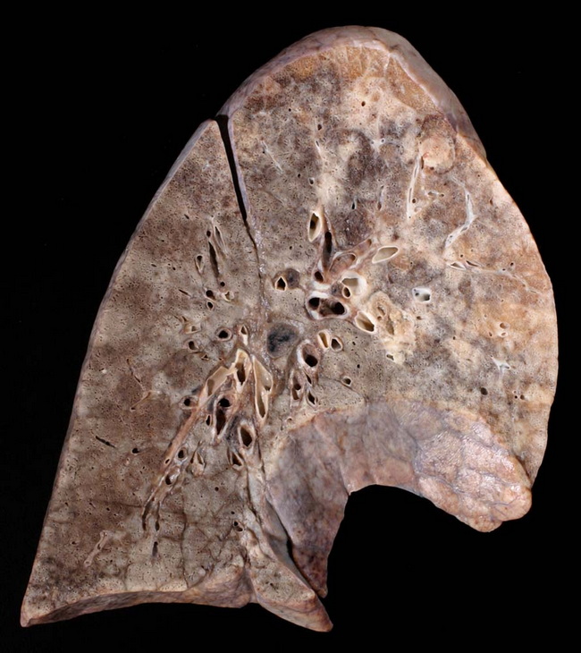
Pneumocystis jirovecii pneumonia with diffuse alveolar damage. The specimen shows edema, congestion and induration of the lung. Microscopically, there was necrotizing pneumonia with evidence of vascular invasive aspergillus and Pneumocystis jirovecii, diffuse alveolar damage with formation of hyaline membranes, and hemorrhage into lung parenchyma. This 65 y/o patient had cryoglobulinemia type III and was hospitalized in a septic state with pancytopenia secondary to immunosuppressive therapy. Image copyright: Pathorama.ch.Pneumocystis jirovecii (previously known as Pneumocystis carinii) is an unusual fungus that was previously considered to be a protozoan parasite. Unlike most fungi, it lacks ergosterol in the cell membrane, does not grow in routine fungal cultures, and responds to trimethoprim-sulphamethoxazole.