
Histoplasmosis : Tongue

slide 3 of 19


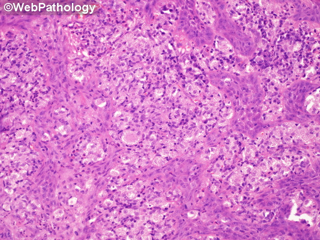
Comments:
Disseminated Histoplasmosis: Section from a tongue ulcer. Numerous epithelioid histiocytes are seen. The irregular squamous epithelial nests are due to tangential sectioning. At this magnification, the causative organism Histoplasma capsulatum can be barely seen as small pink dots within the histiocytes. See higher magnification in the next 3 images. They are more readily seen with PAS or Grocott's stain as 1-3 μm yeast forms.
slide 3 of 19