
Cutaneous Coccidioidomycosis

slide 30 of 52


Comments:
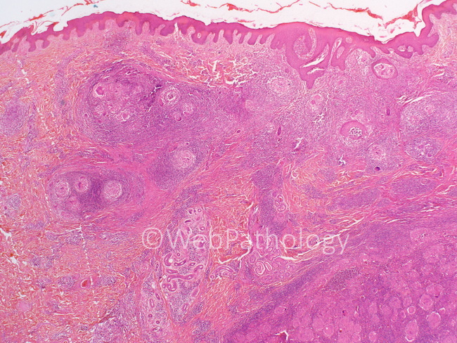
Cutaneous Coccidioidomycosis: Cutaneous lesions show necrotizing and non-necrotizing granulomas in the dermis and subcutaneous tissues. Epithelioid granulomas often have prominent multinucleated giant cells (see next image). Additional findings include pseudoepitheliomatous hyperplasia of the overlying squamous epithelium and a dermal perivascular inflammatory infiltrate. The granulomas are often surrounded by a mixed inflammatory infiltrate of histiocytes, neutrophils, eosinophils, lymphocytes and plasma cells. Eosinophils may be prominent. Eosinophilic abscesses have been reported. The spherules are present within the inflammatory infiltrate or may be seen within the giant cells.
slide 30 of 52