
Pulmonary Coccidioidomycosis

slide 15 of 52


Comments:
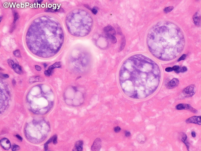
Pulmonary Coccidioidomycosis: In routinely-stained tissue sections, the diagnostic feature is the presence of mature and immature spherules. The background consists of necrotizing and non-necrotizing granulomas and a mixed inflammatory infiltrate of epithelioid histiocytes, neutrophils, eosinophils, lymphoplasmacytic cells, and multinucleated giant cells. Mature spherules are thick-walled, doubly refractile bodies measuring 60-100 μm in diameter and contain multiple endospores measuring 2-5 μm. In addition to mature and immature spherules, mycelia may be found in rare circumstances if the patient develops bronchopulmonary fistula or aerated pulmonary cavities.
slide 15 of 52