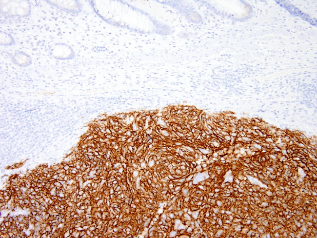
Follicular Dendritic Cell Sarcoma : Immunophenotyping


Comments:
The diagnosis of follicular dendritic cell sarcoma (FDCS) requires positivity for immunohistochemical markers of follicular dendritic cell differentiation combined with negativity for epithelial, soft tissue sarcoma- and melanoma-related markers. The tumor cells in FDCS show markers expressed by normal follicular dendritic cells. They are positive for CD21 (complement receptor type 2), CD23 (Fc fragment of IgE receptor 2), and CD35 (complement receptor type 1; CR1) in almost 95% of cases. The staining may be focal or diffuse and strong. CXCL13, which is diffusely positive, is another highly sensitive and specific marker for FDCS. CXCL13, which belongs to the CXC chemokine family, is selectively chemotactic for B-cells. A recent study found follicular dendritic cell secreted protein (FDCSP) and serglycin (SRGN) to be specific markers for follicular dendritic cells and related tumors. Other positive markers include clusterin, vimentin, fascin, EGFR, and HLA-DR. EMA (but not cytokeratin) is focally positive in 50%-80% of cases. There is variable and weak positivity for CD68. S-100 is positive in about 40% of cases. Ki67 labeling index ranges from 1% to 25%. The small lymphocytes infiltrating the tumor are a mixture of mostly T-cells and a few B-cells. Negative markers include: CD45/CD45RB, CD20, CD79a, CD3, CD34, CD30, CD1a, lysozyme, myeloperoxidase, HMB-45, desmin, muscle-specific actin, and high molecular weight cytokeratins. The image shows FDCS arising in the colon. The tumor was strongly positive for CD23. Image courtesy of: ARP Press; used with permission.


