
Sezary Syndrome : Microscopic Features
Home
 Hematopathology
Mature T-cell & NK-cell Neoplasms
Mycosis Fungoides & Sezary Syndrome
Sezary Syndrome : Microscopic Features
Hematopathology
Mature T-cell & NK-cell Neoplasms
Mycosis Fungoides & Sezary Syndrome
Sezary Syndrome : Microscopic Features

slide 70 of 73


Comments:
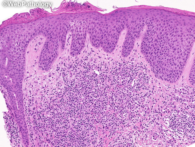
Sezary syndrome (SS) - Microscopic Features: In SS, the findings in diagnostic skin biopsies are similar to the late patch- or plaque stage of mycosis fungoides (MF). There is a dense band-like lymphocytic infiltrate in the dermis. Epidermotropism is seen in 20%-40% of cases and is less prominent. Small uniform lymphocytes are the predominant cell population. Large atypical cells with cerebriform nuclei (Sezary cells) are in the minority but their presence is helpful in making the diagnosis of SS in the appropriate clinical setting. In a small percentage of cases, the dermal infiltrate is composed entirely of large atypical cells resembling a large cell lymphoma.
slide 70 of 73