
SPTCL : Differential Diagnosis


Comments:
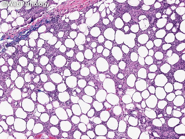
Differential Diagnosis of Subcutaneous Panniculitis-like T-cell Lymphoma (SPTCL): Lupus erythematosus panniculitis (LEP): LEP and SPTCL share morphologic similarities and may represent the two ends of the spectrum of the same disease process. The features supporting the diagnosis of LEP include: numerous plasma cells, areas of hyalinization, presence of CD123+ plasmacytoid dendritic cells, nodular aggregates of B-cells with reactive germinal centers, low Ki-67 labeling index, and lack of clonal rearrangement of TCR genes. Mycosis fungoides (MF): Rarely, MF may show subcutaneous lesions; however, the neoplastic cells have CD4+ phenotype, in contrast to the CD8+ cells of SPTCL. Cutaneous gamma-delta T-cell Lymphoma (GDTCL): Features favoring GDTCL include dermal or epidermal involvement with prominent epidermotropism. It is usually CD4-, CD8-, Beta F1-, CD56+, and gamma-delta T-cell receptor+. Cutaneous extranodal NK/T-cell lymphoma, nasal type: It shows marked involvement of dermis and is CD56+, EBV+, and lacks monoclonal rearrangement of TCR genes. Subcutaneous Anaplastic Large Cell Lymphoma: It shows large pleomorphic CD30+ and CD4+ cells in the infiltrate. The image shows neoplastic lymphocytes infiltrating subcutaneous fat in a case of SPTCL. The pattern resembles lobular panniculitis.