
EATL : Microscopic


Comments:
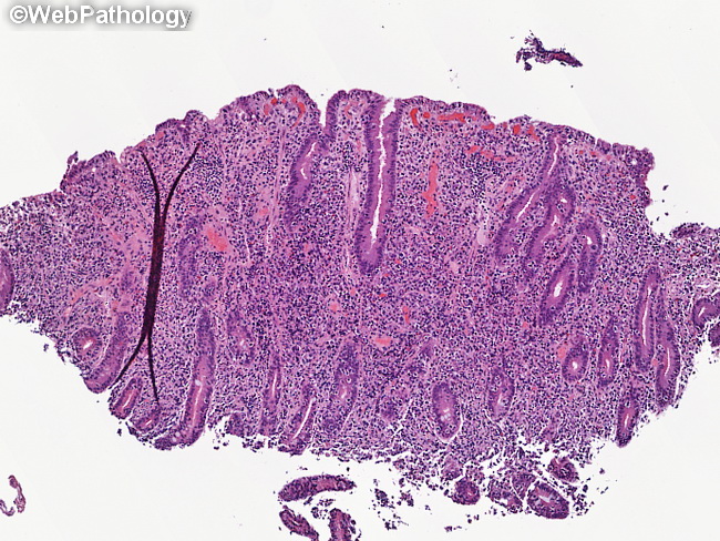
Microscopic Features of Enteropathy-associated T-cell Lymphoma: The involved segment of the bowel is frequently ulcerated. The adjacent mucosa may be normal or show features of celiac disease (as seen here), including villous atrophy, crypt hyperplasia, intraepithelial lymphocytosis, and increased numbers of plasma cells and lymphocytes in the lamina propria. The neoplastic T-cells form a dense infiltrate with destruction of the native architecture. They are moderately pleomorphic, medium-to-large size with round or angulated nuclei and prominent nucleoli. About 40% of cases show large cell or anaplastic phenotype. The tumor cells display angiocentric and angioinvasive pattern with extensive necrosis. There is a prominent component of inflammatory cells, including histiocytes and eosinophils. The mesenteric lymph nodes are frequently involved and show intrasinusoidal or paracortical infiltration by neoplastic cells. Some lymph nodes simply show necrosis or cavitation (and replacement with lymph fluid) in the absence of a recognizable lymphoma.