
Cutaneous Marginal Zone Lymphoma : Morphology


Comments:
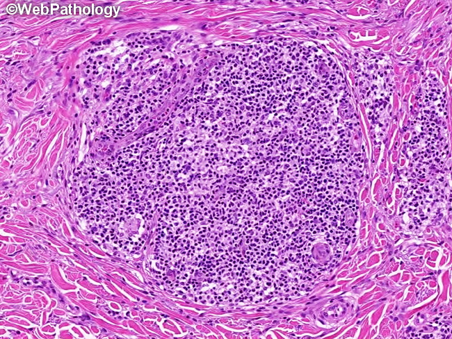
Cutaneous Marginal Zone Lymphoma (CMZL) - Morphology (continued) : The infiltrate contains reactive lymphoid follicles with germinal centers and mantle zones. They are surrounded by centrocyte-like marginal zone cells with slightly irregular nuclei, inconspicuous nucleoli, and pale or clear cytoplasm. In addition to marginal zone cells, there are small lymphocytes, lymphoplasmacytoid cells, and plasma cells, admixed with centroblast-like or immunoblast-like cells and reactive T-cells. In most cases of CMZL, the neoplastic marginal zone cells form a minority of the infiltrate. In rare cases, the neoplastic marginal zone cells infiltrate into the germinal centers (follicular colonization) as seen here. Lymphoepithelial lesions are uncommon and, when present, affect hair follicles and sweat glands. Mitotic activity is low.