
Gastric MALT Lymphoma - Morphology


Comments:
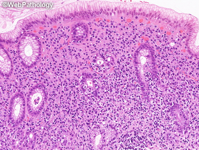
Gastric MALT Lymphoma - Morphology: At low magnification, the gastric biopsies show a dense, expansile monotonous lymphoid infiltrate deep within the mucosa (as seen in the previous two images). The infiltrate consists of monomorphic small lymphocytes with clear cytoplasm (which creates pericellular halos or clearing). Lymphoepithelial lesions, consisting of three or more neoplastic marginal zone lymphocytes, invade the glandular epithelium and distort the glandular architecture. This is often accompanied by eosinophilic degeneration of the epithelium. Both muscularis mucosae and propria may be invaded and splayed by the lymphoid cells. Plasma cell differentiation is quite common (about one-third of cases) and is pronounced beneath the surface gastric epithelium. This gastric biopsy with MALT lymphoma shows several lymphoepithelial lesions and prominent plasmacytic differentiation.