
Ganglion : Differential Diagnosis

slide 10 of 11


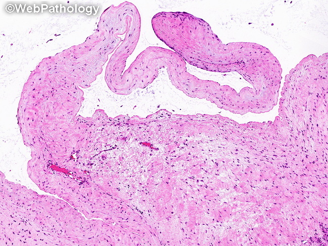
Comments:
Differential Diagnosis of Ganglion: Myxoma: The initial stages of ganglion may just show focal myxoid changes which may be confused with a myxoma. However, ganglion most often occurs at a characteristic location (dorsum of wrist) and consists of multilocular thick-walled cystic spaces in addition to the myxoid areas. Synovial (Baker) cysts: They are formed by herniation of synovial membrane through the weakened joint capsule. They are lined by synovium and communicate with the joint space. Both of those features are absent in ganglion. The image shows thick fibrous wall of a ganglion. Note the myxoid areas in the stroma.
slide 10 of 11