
Syringocystadenoma Papilliferum

slide 65 of 126


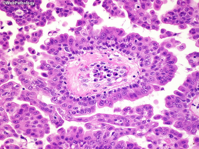
Comments:
The above photomicrograph shows a cluster of plasma cells within the stroma of a papillary frond. This is a helpful feature for distinguishing SP from hidradenoma papilliferum. Treatment options for SP include surgical excision, CO2 laser excision, and Mohs micrographic surgery.
slide 65 of 126