
Metanephric Adenoma : References


Comments:
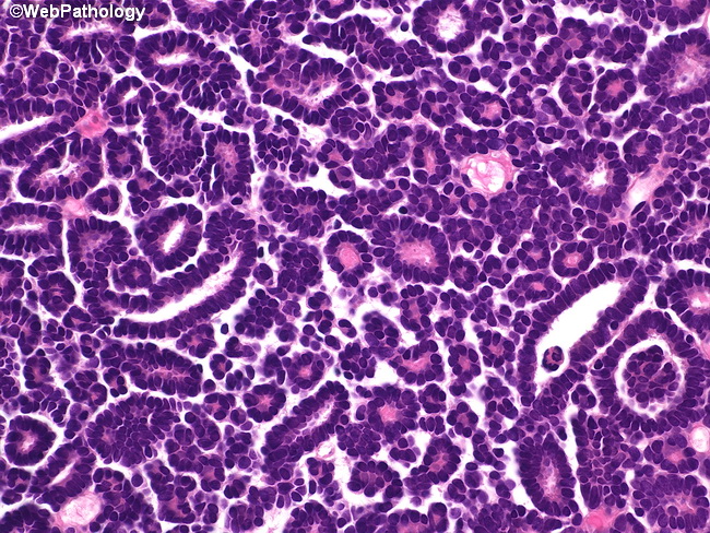
References: 1. Goldblum, J. R. et al (2018). Rosai and Ackerman's Surgical Pathology - Eleventh Edition. Philadelphia, PA. Elsevier. 2. Amin MB et al (2010). Diagnostic Pathology: Genitourinary. Amirsys Publishing Inc.3. Cheng, L., MacLennan G. T., & Bostwick, D. G. (2020). Urologic Surgical Pathology - 4th Edition. Philadelphia, PA. Elsevier. 4. WHO Classification of Tumours Editorial Board. Urinary and male genital tumours [Internet]. Lyon (France): International Agency for Research on Cancer; 2022 [cited 2022 April 20]. (WHO classification of tumours series, 5th ed.; vol. 8). Available from: https://tumourclassification.iarc.who.int/chapters/36.5. Kinney SN et al. Metanephric adenoma: the utility of immunohistochemical and cytogenetic analyses in differential diagnosis, including solid variant papillary renal cell carcinoma and epithelial-predominant nephroblastoma. Modern Pathology (2015) 28, 1236-1248. About this image: Metanephric adenoma composed of small, uniform, round acini and tubular structures separated by scant stroma. The lining epithelial cells are uniform with hyperchromatic nuclei and have scant cytoplasm.