
Psammomatous Meningioma

slide 36 of 60


Comments:
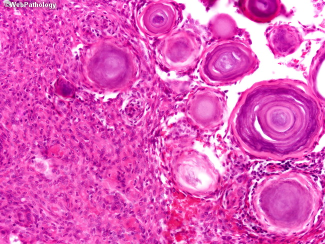
The picture illustrates two Grade I patterns coexisting in the same tumor, perhaps representing different stages in the progression of this tumor. On the left, sheets of meningothelial cells of a probable transitional meningioma are noted. On the right we can observe psammoma bodies exhibiting different ages based on their level of calcification.
slide 36 of 60