
Fibrous Meningioma

slide 22 of 60


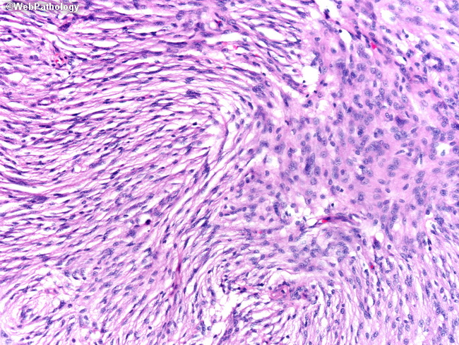
Comments:
High power view of the previous photomicrograph. Benign-looking fibroblastic meningioma with monomorphic elongated cells and spindly nuclei. At the right upper corner a fascicle has been cut horizontally. In this field the bland cytology of meningothelial cells is better appreciated. The differential diagnosis of fibroblastic meningioma includes schwannoma and solitary fibrous tumor. Schwannomas show strong diffuse positivity for S-100 and solitary fibrous tumor are immunoreactive for CD34.
slide 22 of 60