
Parathyroid Adenoma
slide 1 of 22


Comments:
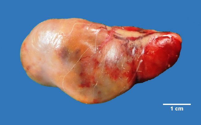
This parathyroid adenoma was removed from a 39 y/o female who presented with abdominal pain. During the work-up, a CT scan demonstrated a well-defined, hypodense mildly enhancing lesion behind the lower pole of the left lobe of the thyroid. Lytic lesions were noted in the humerus and one of the ribs. Serum calcium was 12 mg/dl and serum parathormone levels were 2275 pg/ml (ref. range 12 to 72 pg/ml). The tumor measured 5 cm in greatest dimension and was surrounded by a thin fibrous capsule. Contributed by : Dr. Sanjay D. Deshmukh, Department of Pathology, Smt. Kashibai Navale Medical College, Pune, India.
slide 1 of 22