
Luteinized Thecoma : Gross & Micro


Comments:
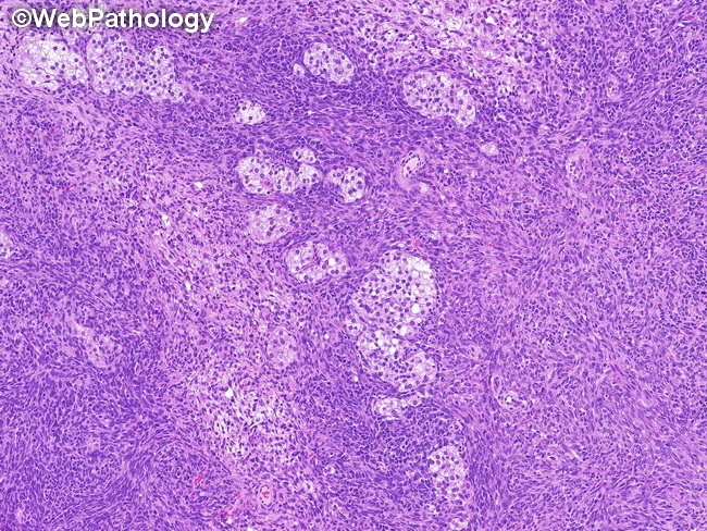
Gross Pathology: Luteinized thecomas are grossly similar to typical thecomas. They are unilateral, solid yellow masses which may have areas of cystic change and calcification. Luteinized thecomas with sclerosing peritonitis (LTSP) are almost always bilateral and cause variable enlargement of ovaries which may develop a cerebriform appearance. The cut surface shows cortical edema and cystic changes without a discrete mass. Microscopic Appearance: Luteinized thecomas resemble typical thecomas but contain lutein (steroid) cells, singly or in clusters. The lutein cells stand out sharply due to their abundant light pink cytoplasm against a basophilic background of typical thecoma. They have round vesicular nuclei with single prominent nucleoli. There is no nuclear atypia but mitotic activity may be brisk.