
Ovarian Fibroma : Differential Diagnosis


Comments:
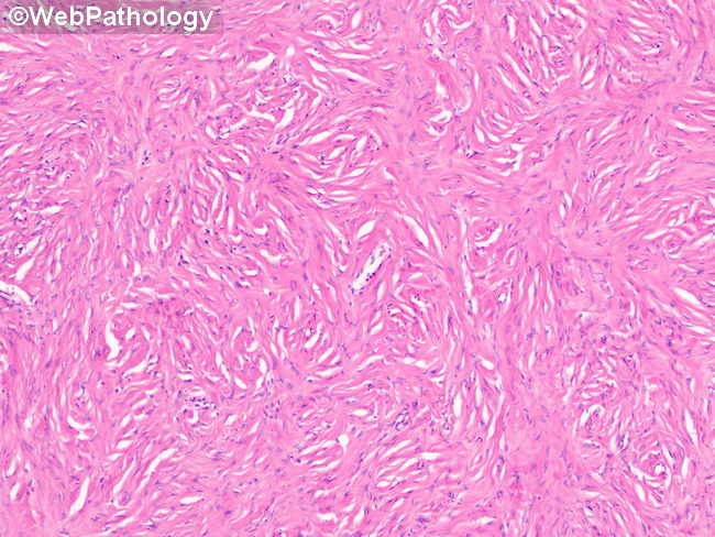
Differential Diagnosis of Ovarian Fibroma (continued from the previous image): Thecoma: Fibroma and thecoma have overlapping morphology and the two may coexist within the same tumor (fibrothecoma). Grossly, thecomas are often yellow rather than white. They are more often associated with evidence of hormone production (e.g. endometrial hyperplasia or carcinoma). Thecomas have nodular architecture and show polygonal or fusiform cells with abundant pale or vacuolated cytoplasm. They are positive for inhibin and CD10 (which may be focal). These features are lacking in fibromas, which are negative (or weakly +ve) for CD10 and show patchy positivity for inhibin. Fibrosarcoma: Fibrosarcomas of the ovary are quite rare and tend to present in post-menopausal women as large, bulky abdominal masses with frequent hemorrhage and necrosis. Extra-ovarian involvement may be seen. Microscopically, they resemble cellular fibromas. Presence of moderate to severe cytologic atypia and atypical mitotic figures favors fibrosarcoma over cellular or mitotically-active fibroma. (differential diagnosis continues in the next image) This image of an ovarian fibroma shows short fascicles of bland spindle cells with a storiform pattern in abundant collagenous stroma.