
Mature Cystic Teratoma

slide 63 of 183


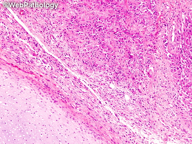
Comments:
Mature Cystic Teratoma of the Ovary: The image shows cartilage on the lower left and neuroglial tissue on the upper right.
slide 63 of 183