
Papillary Urothelial Neoplasm of LMP
Home
 Genitourinary
Urinary Bladder
Papillary Urothelial Neoplasms I
Papillary Urothelial Neoplasm of LMP
Genitourinary
Urinary Bladder
Papillary Urothelial Neoplasms I
Papillary Urothelial Neoplasm of LMP

slide 10 of 30


Comments:
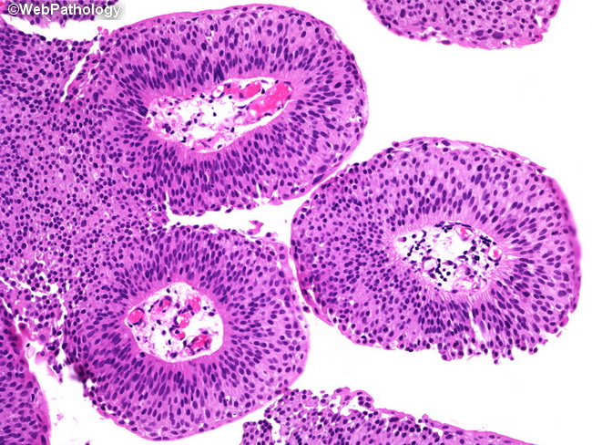
Higher magnification of the previous image. Note the orderly arrangement of urothelial cells with no loss of polarity. The nuclei are crowded and appear minimally enlarged. Mitotic figures are not seen. When present, they are confined to the basal layers. Nucleoli are not prominent.
slide 10 of 30