
Giant Cell Arteritis : Microscopic Features

slide 9 of 17


Comments:
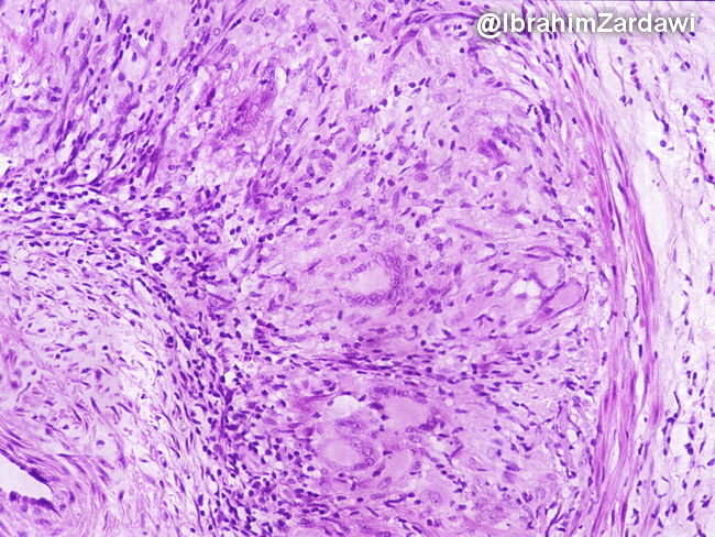
Microscopic Features: The inflammatory infiltrate in giant cell arteritis is present adjacent to the internal elastic lamina. It consists of lymphocytes, plasma cells, epithelioid histiocytes and multinucleated giant cells which often contain phagocytosed fragments of internal elastic lamina. Several giant cells are present in this image. However, giant cells are often absent and not required for diagnosis. Medial necrosis is minimal to absent and there are no compact well-formed granulomas. Image courtesy of: Dr. Ibrahim Zardawi; used with permission.
slide 9 of 17