
Giant Cell Arteritis : Microscopic Features

slide 8 of 17


Comments:
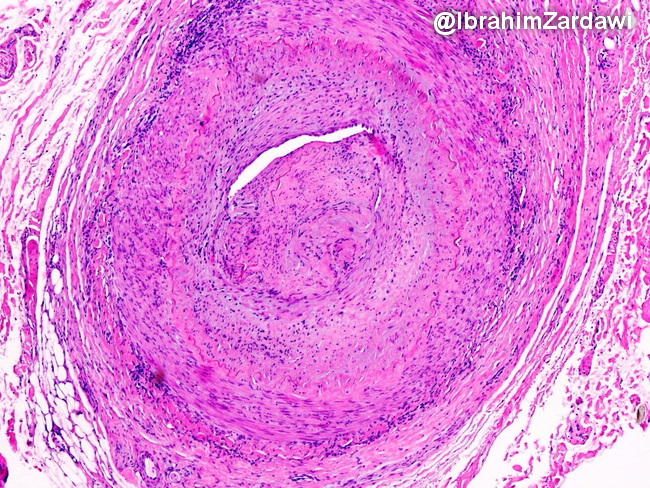
Microscopic Features: Giant cell arteritis shows transmural (full thickness) granulomatous inflammation with segmental disruption of internal elastic lamina. Perivascular inflammation alone without involvement of all layers (intima, media, and adventitia) is not diagnostic. Inflammatory changes extend to the small vessels (vasa vasorum, venules) in the tunica adventitia of the involved artery. In addition to the transmural inflammation, this low magnification view also shows severe intimal hyperplasia causing near-total occlusion of the lumen. Image courtesy of: Dr. Ibrahim Zardawi; used with permission.
slide 8 of 17