
Giant Cell Arteritis : Temporal Artery Biopsy


Comments:
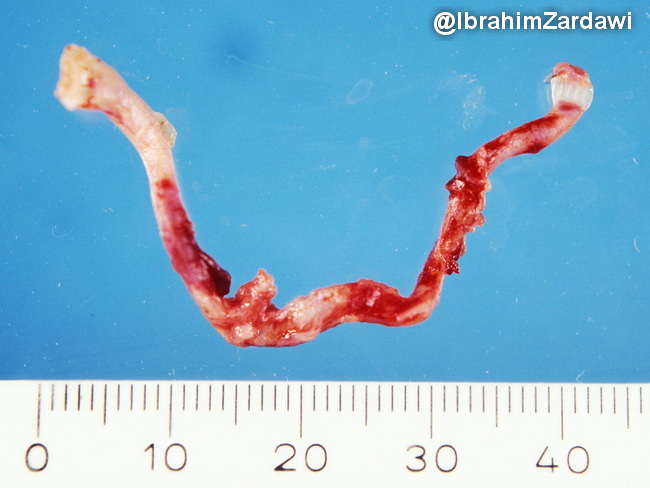
Superficial temporal artery biopsy (TAB): It is the gold standard for the diagnosis of giant cell arteritis (GCA), with a >90% positive predictive value. TAB is indicated when a patient presents with symptoms and signs suggestive of GCA (patient >50 yrs, new-onset headache, visual changes, jaw claudication, elevated ESR and CRP). Grossly, the TAB specimen may show segmental wall thickening with nodular appearance and lumen narrowing. Since complications like permanent blindness can develop, treatment with corticosteroids should not be delayed until after the TAB is done. Up to 10 days of corticosteroid therapy has no effect on biopsy findings. Diagnostic yield of TAB decreases after 30 days of corticosteroid therapy.Some advocate processing the biopsied vessel whole so that it firms up for serial sectioning and embedding. Inflammatory changes are commonly segmental with skip areas. Therefore, exhaustive serial sectioning and submission of multiple cross sections is necessary. A negative biopsy (about 40% of cases) does not exclude the diagnosis of GCA. Image courtesy of: Dr. Ibrahim Zardawi; used with permission.