
Cardiac Fibroma : Gross Pathology


Comments:
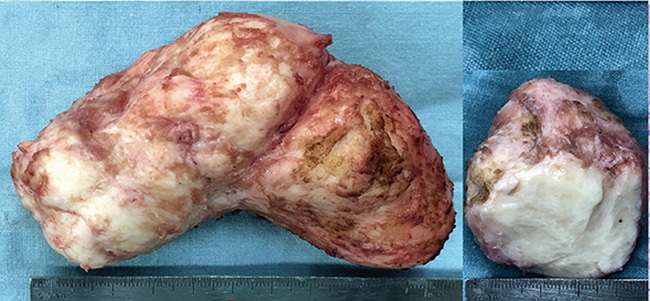
Gross Pathology: Cardiac fibromas form a solitary, circumscribed, bulging, firm mass that can reach massive size (often > 10 cm). The cut surface is grey-white with a whorled appearance, resembling uterine fibroids. They are often calcified which can be detected on chest x-ray and helps distinguish them from rhabdomyomas which rarely calcify. The image shows a 12 cm cardiac rhabdomyoma resected from the posterolateral wall of left ventricle. The patient was a 7.5 y/o male who presented with sustained ventricular tachycardia. Image Source: Qian T et al. Surgery for Primary Cardiac Tumors in Children: Successful Management of Large Fibromas. Frontiers in Cardiovascular Medicine, March 2022; Vol. 9, Article 808394; image cropped and used under Creative Commons Attribution License.