
Cardiac Fibroma : Clinical Features


Comments:
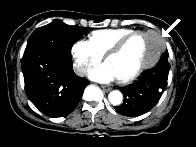
Clinical Features: Cardiac fibromas often involve the left ventricle (usually interventricular septum) followed by right ventricle. Atrial location is rare. Depending upon their size and location, they can cause arrhythmias (two-thirds of patients), cardiac failure, or even sudden death. They can engulf coronary arteries and compromise cardiac blood supply.Case History: This chest CT with contrast is from a 40 y/o male who presented with chest pain. An intramural mass is seen originating from the left ventricular free wall. The mass is isodense to the ventricular wall and contained calcification. The diagnosis of cardiac fibroma was confirmed on histopathologic examination of the resected mass. Case courtesy of Azza Elgendy, Radiopaedia.org. From the case rID: 38974