
Cardiac Rhabdomyoma : Intro & Radiology


Comments:
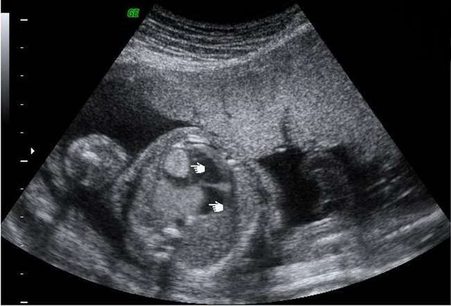
Rhabdomyoma is the most common cardiac tumor in children (followed by fibromas) and usually occurs as multiple nodules (95% of cases). It is often associated with tuberous sclerosis complex (60-80% of cases). It arises from cardiac myocytes and may represent a hamartomatous lesion rather than a true neoplasm.
Echocardiography is the primary diagnostic modality for evaluation of cardiac tumors in children. Cardiac rhabdomyomas usually appear as multiple, round, homogenous, hyperechogenic, intramural or intracavitary masses. The image shows a fetal cardiac rhabdomyoma discovered on antenatal scan at 20 weeks gestation. Multiple echogenic lesions are seen in the left atrioventricular region. Case courtesy of Dr Effendi Mansoor, Radiopaedia.org. From the case rID: 17302