
Cardiac Sarcoidosis : Differential


Comments:
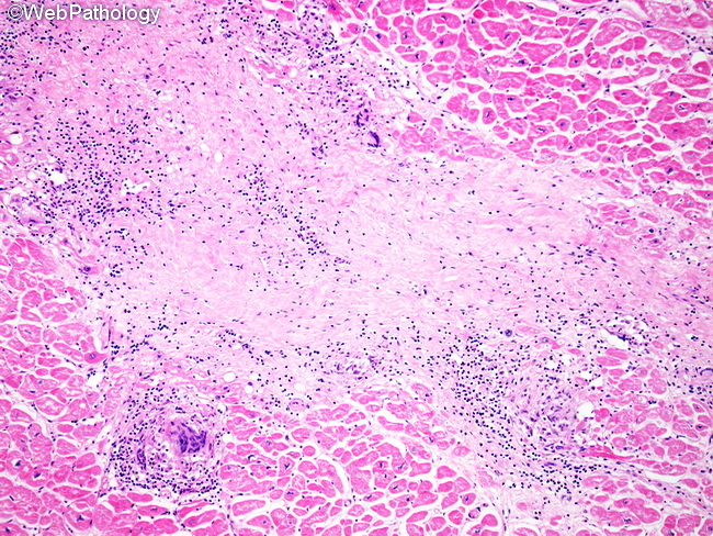
The differential diagnosis of cardiac sarcoidosis includes the following: Giant Cell Myocarditis: This is a rapidly progressive disease which can quickly lead to congestive heart failure and death within weeks of onset. During the active phase, histologic findings include myocyte necrosis, multinucleated giant cells, and an inflammatory infiltrate of CD8+ T-cells, macrophages, and eosinophils. Granulomas and significant fibrosis are not seen. Lymphocytic Myocarditis: This is usually seen after a viral infection (most commonly respiratory). Endomyocardial biopsies show patchy lymphocytic infiltrates and myocyte destruction. The inflammation subsides with considerable scarring and cardiomyopathy which may require transplant. Allergic (hypersensitivity) Myocarditis: This may occur as a result of allergic reaction to drugs or other ingested compounds. Onset is rapid and there is precipitous decline of cardiac function with heart failure. Endomyocardial biopsies show a prominent eosinophilic infiltrate. Tuberculous Myocarditis: It is quite rare and may be seen in patients with known tuberculosis. Biopsies show caseating granulomas containing acid-fast bacilli. The image shows a scar-like area of interstitial fibrosis with multinucleated giant cells and lymphocytic infiltrate.