
Inclusion Body Fibromatosis : Electron Microscopy
Home
 Soft Tissue
Fibroblastic
Fibrous Tumors of Infancy
Inclusion Body Fibromatosis : Electron Microscopy
Soft Tissue
Fibroblastic
Fibrous Tumors of Infancy
Inclusion Body Fibromatosis : Electron Microscopy

slide 59 of 60


Comments:
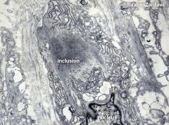
Inclusion Body Fibromatosis - Ultrastructure: With electron microscopy, the inclusion bodies appear to be non-membrane bound tight bundles of actin microfilaments. Image courtesy of: Dr. Ibrahim Zardawi; used with permission.
slide 59 of 60