
Inclusion Body Fibromatosis : Morphology

slide 55 of 60


Comments:
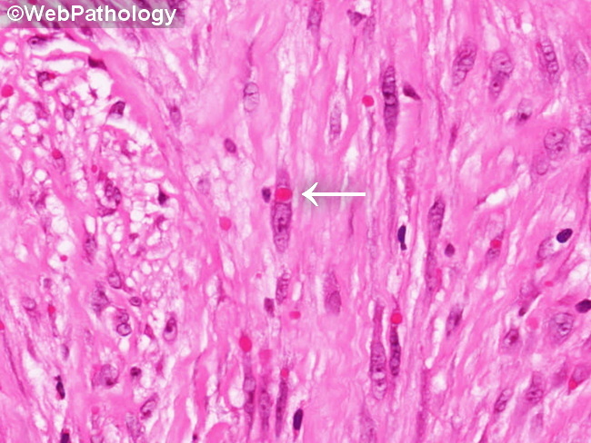
Inclusion Body Fibromatosis: The center of the image shows a prominent eosinophilic cytoplasmic inclusion adjacent to the nucleus within a spindle cell (arrow). Numerous additional inclusions can also be seen throughout the field. The inclusions resemble red blood cells but can be easily distinguished from them by their intracellular location and variability in size. The spindle cells have pale eosinophilic cytoplasm and elongated nuclei. There is no cytologic atypia and mitotic activity is not increased.
slide 55 of 60