
Inclusion Body Fibromatosis : Morphology

slide 54 of 60


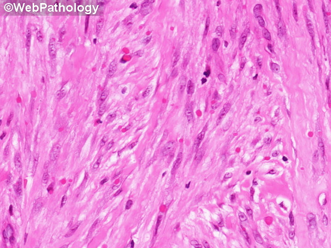
Comments:
Microscopic Features of Inclusion Body Fibromatosis (continued from the previous image): The most characteristic finding, that gives the tumor its name, is the presence of variable numbers of small round eosinophilic cytoplasmic inclusions close to the nucleus within spindle cells. They resemble red blood cells but are more variable in size (as nicely shown here). The inclusions may be numerous and easy to find or few in number and hard to detect in some cases on routine H&E stains. With Masson trichrome, they stain deep red and are somewhat easier to visualize. They don't stain with Alcian blue, colloidal iron, or PAS stains.
slide 54 of 60