
Inclusion Body Fibromatosis : Morphology

slide 53 of 60


Comments:
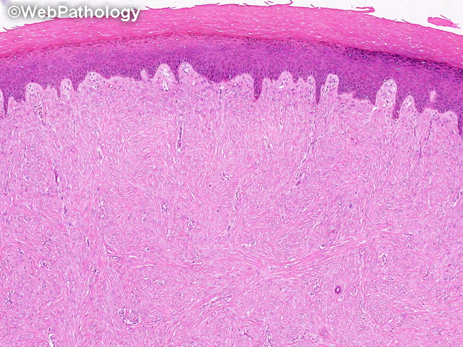
Gross Pathology: The resected specimens are small, skin-covered solid nodules with a whitish-grey firm cut surface. Microscopic Features: The lesion is poorly circumscribed and extends into deep dermis and subcutis and surrounds the dermal appendages. The overlying epidermis may show acanthosis or hyperkeratosis. The lesion consists of a uniform population of bland myofibroblastic cells in a dense collagenous stroma. microscopic features continue in the next image
slide 53 of 60