
Calcifying Aponeurotic Fibroma : Differential Diagnosis


Comments:
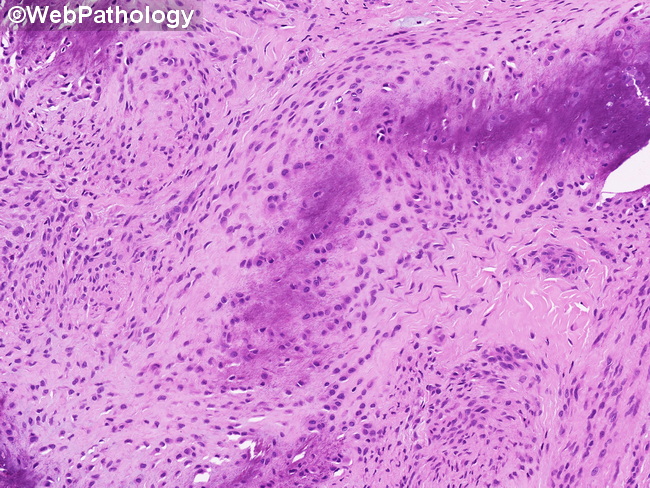
Differential Diagnosis: The differential diagnosis of calcifying aponeurotic fibroma (CAF) depends upon the patient's age and includes the following: Infantile Fibromatosis (IF): Early lesions of CAF (usually in infants and small children) are ill-defined and lack calcification and may be mistaken for IF. Features favoring IF include: location in the head and neck or extremities, elongated fibroblasts in a myxoid background, absence of calcification and cartilaginous metaplasia, and absence of giant cells. Palmar and Plantar Fibromatoses: They are uncommon in children. The lesions are circumscribed with a nodular appearance and lack calcification and cartilaginous areas. Monophasic (fibrous type) Synovial Sarcoma: CAF with a prominent spindle cell component can mimic monophasic synovial sarcoma. Presence of epithelial marker immunoreactivity and SS18 gene aberrations favor synovial sarcoma. Soft Part Chondroma (SPC): Both CAF and SPC are common in hands; however, SPC occurs in older adults and has a lower recurrence rate. Features favoring SPC over CAF include: well-circumscribed lobulated mass composed predominantly of well-developed chondroid areas, high cellularity, and lack of plump myofibroblastic cells. Acral Fibroblastic Spindle Cell Neoplasm: This is a recently described fibroblastic tumor with hyalinization and stippled calcification. However, it is seen in adults and carries EWSR1-SMAD3 gene fusions. Rheumatoid nodule and even schwannoma enter into the differential diagnosis.