
Calcifying Aponeurotic Fibroma : Microscopic
Home
 Soft Tissue
Fibroblastic
Fibrous Tumors of Infancy
Calcifying Aponeurotic Fibroma : Microscopic
Soft Tissue
Fibroblastic
Fibrous Tumors of Infancy
Calcifying Aponeurotic Fibroma : Microscopic

slide 24 of 60


Comments:
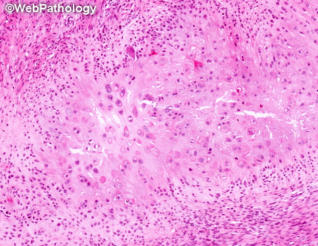
Microscopic Features of Calcifying Aponeurotic Fibroma (continued from the previous image): The center of the image shows chondrocyte-like cells in hyalinized and partly calcified matrix. It is surrounded by radiating and swirling arrays of plump fibroblastic/myofibroblastic cells. Calcification and chondroid metaplasia are more readily seen in lesions removed from older children and young adults. Immunohistochemistry: Immunostains for SMA, MSA and CD99 are positive in the fibroblastic areas. S100 protein is positive in chondroid foci. Nuclear β-catenin is not seen.
slide 24 of 60