
Calcifying Aponeurotic Fibroma : Microscopic
Home
 Soft Tissue
Fibroblastic
Fibrous Tumors of Infancy
Calcifying Aponeurotic Fibroma : Microscopic
Soft Tissue
Fibroblastic
Fibrous Tumors of Infancy
Calcifying Aponeurotic Fibroma : Microscopic

slide 23 of 60


Comments:
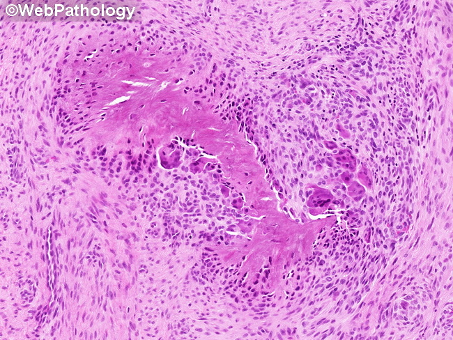
Microscopic Features of Calcifying Aponeurotic Fibroma (continued from the previous image): Areas of calcification are better defined in lesions removed from older children and young adults and range from small granules to large amorphous basophilic deposits. As the calcified areas mature, they are surrounded by circumferential or radiating arrays of chondrocyte-like cells and plump fibroblasts/myofibroblasts. Osteoclast-type giant cells are also commonly seen (as depicted here). Uncommonly, there are ossified foci with extramedullary hematopoiesis.
slide 23 of 60