
Calcifying Aponeurotic Fibroma : Clinical Features


Comments:
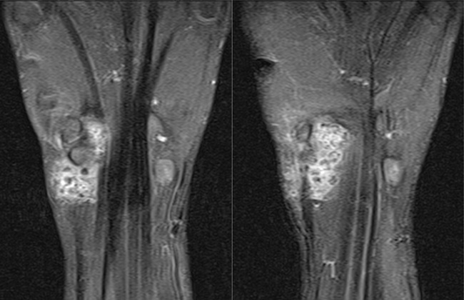
Clinical Presentation: Calcifying aponeurotic fibroma (CAF) makes up 1-2% of all fibrous tumors of childhood. It presents as a small, painless, slow-growing, nodular or ill-defined mass in hands (palmar surface of hands and fingers) and feet (plantar surface of feet and toes). Wrists and ankle are less commonly involved. Rare sites include elbow, upper arm, scalp, trunk, and gluteal region. The age range is 5 to 15 years with a male predilection. Rare cases have been reported in adults. It is usually asymptomatic but may cause mild tenderness in some cases. On examination, it appears as a poorly-defined subcutaneous mass, often attached to tendons, aponeurosis, or fascia. Imaging: This MRI of hand (coronal, T1 C+) is from a 13 y/o male who presented with a painless mass on the palmar aspect of his right wrist of long-duration and not causing limitation of movement. It shows a poorly-encapsulated soft tissue mass with intense heterogenous enhancement with contrast. The darker intralesional nodules are calcific foci. The diagnosis of CAF was confirmed on histology. Case courtesy of Hrishikesh VJ, Radiopaedia.org. From the case rID: 88423