
Myxofibrosarcoma : Morphology


Comments:
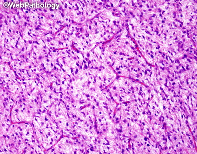
Gross Pathology: Superficially located myxofibrosarcomas consist of multiple nodules with variably gelatinous or mucoid cut surface. Deep-seated tumors are usually firm with infiltrative margins. Microscopic Features: Myxofibrosarcomas have variable amounts of myxoid stroma and cellular components. By definition, the myxoid areas form >50% of the tumor. The myxoid foci may gradually blend into the cellular areas or the two components may be sharply demarcated. The myxoid areas have curvilinear vessels (as clearly seen in this image) and less prominent storiform pattern. The tumor cells and inflammatory cells condense around the vessels. The tumor cells may be spindled or epithelioid. Epithelioid cells may ingest stromal mucin and have bubbly cytoplasm resembling physaliferous cells of chordoma.