
Dedifferentiated Liposarcoma : Case History


Comments:
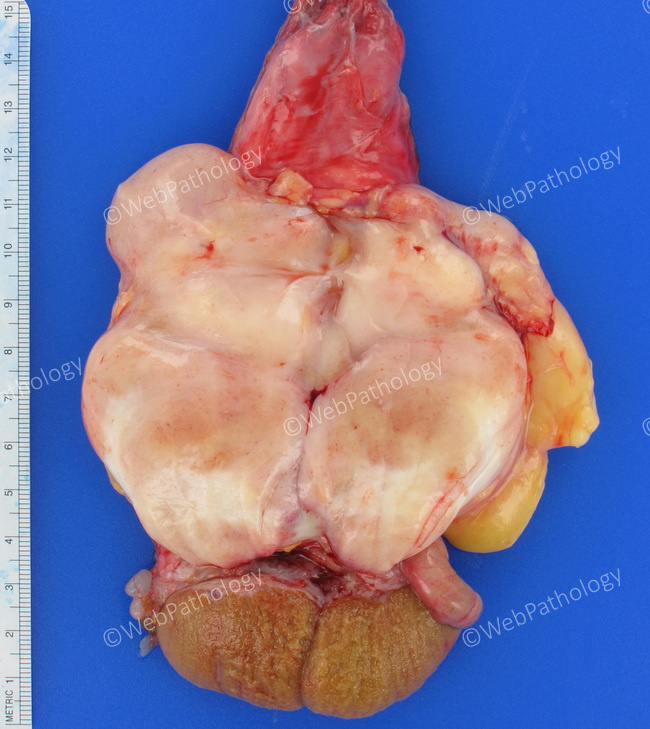
Gross Pathology: This radical orchiectomy specimen is from a 75 y/o male who presented with painless enlargement of right hemiscrotum. The patient reported that the mass had become firmer in the last 4-5 months. Grossly, there was a 10 cm lobulated, pink-tan mass centered in the spermatic cord. It had firm consistency but was punctuated by scattered hard, nodular areas. The testis proper was free of lesions. The microscopic appearance was that of a well-differentiated sclerosing liposarcoma (WDL) admixed with more cellular spindle cell areas (dedifferentiated foci) showing considerable nuclear hyperchromasia and pleomorphism. MDM2 amplification by FISH was positive confirming the diagnosis of dedifferentiated liposarcoma arising in a WDL.