
Atypical Lipomatous Tumor/Well-Diff Liposarcoma : Differential


Comments:
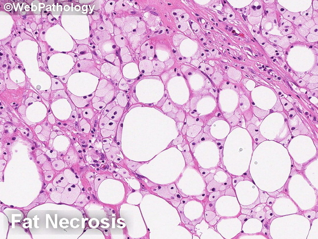
Differential diagnosis of atypical lipomatous tumor/well-differentiated liposarcoma (continued):Fat Necrosis: Areas of fat necrosis show adipocyte drop out, chronic inflammation and lipid-laden macrophages with finely granular or microvacuolated cytoplasm. In contrast to lipoblasts, the macrophages are smaller with more uniform shape. They have small, round, regular nucleus that is not indented by the evenly dispersed cytoplasmic vacuoles. Fat Atrophy in Starvation/Malnutrition: There is loss of intracellular lipid resulting in cell shrinkage. The nucleus becomes relatively prominent and the adipocytes may resemble lipoblasts. An important diagnostic clue is that the adipocytes are of uniform size and maintain lobular architecture. In advanced cases, there is lipofuscin pigment in the cytoplasm.