
Atypical Lipomatous Tumor/Well-Diff Liposarcoma : Imaging
Home
 Soft Tissue
Lipomatous
Liposarcoma (Well-diff. & De-Diff)
Atypical Lipomatous Tumor/Well-Diff Liposarcoma : Imaging
Soft Tissue
Lipomatous
Liposarcoma (Well-diff. & De-Diff)
Atypical Lipomatous Tumor/Well-Diff Liposarcoma : Imaging

slide 13 of 85


Comments:
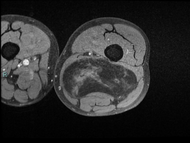
Atypical Lipomatous Tumor/Well-differentiated Liposarcoma - Imaging: This MRI (axial T1 fat sat image) is from a 70 y/o male with a mass in posterior distal left thigh. In the posterior compartment of the left thigh, there is a well-circumscribed mass with a main adipose component and a central irregular solid component. It displays contrast enhancement. The neurovascular bundle and femur are not infiltrated. The diagnosis of ALT/WDL was confirmed on histologic examination. Case courtesy of Domenico Nicoletti, Radiopaedia.org. From the case rID: 45044
slide 13 of 85