
Hibernoma : Microscopic Features

slide 7 of 27


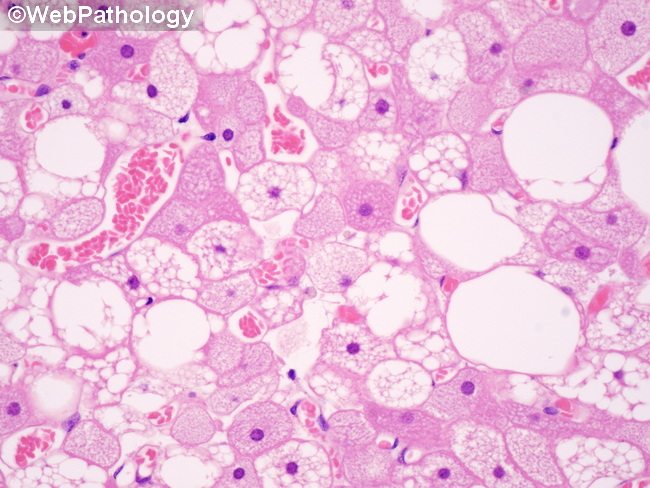
Comments:
Microscopic Features: In a classic hibernoma, greater than 70% of the tumor resembles brown fat. It shows an admixture of: 1) large polygonal, round or ovoid cells with abundant granular eosinophilic cytoplasm, distinct cytoplasmic borders, and round centrally-placed nuclei with prominent nucleoli; 2) multivacuolated large adipocytes with central nuclei; and 3) variable numbers of mature univacuolated adipocytes (white fat). All three cell types are seen in this image. There is no cytologic atypia and mitotic activity is not increased.
slide 7 of 27