
Hibernoma : Locations

slide 3 of 27


Comments:
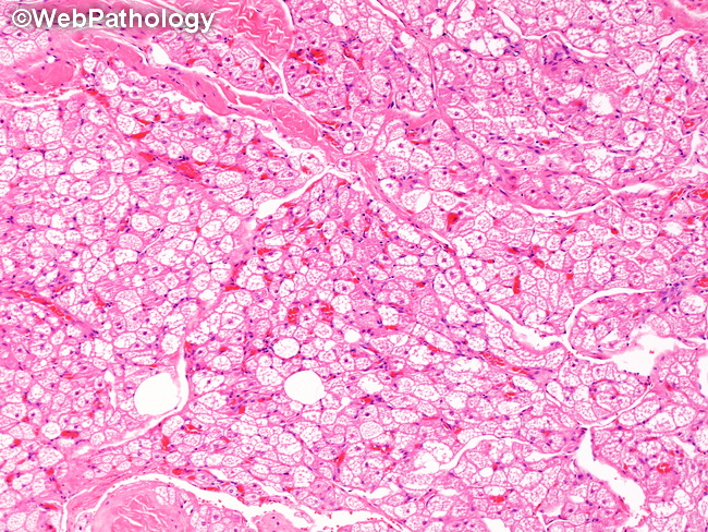
Locations: Hibernomas usually arise as subcutaneous masses in the back (interscapular and scapular region), thigh, axilla, neck, chest, and arms. Tumors in abdomen/retroperitoneum/mediastinum comprise <10% of cases. Myxoid and spindle cell subtypes occur in the posterior neck and shoulder. About 20% of cases are intramuscular.
slide 3 of 27