
Chondroid Lipoma : Gross Pathology


Comments:
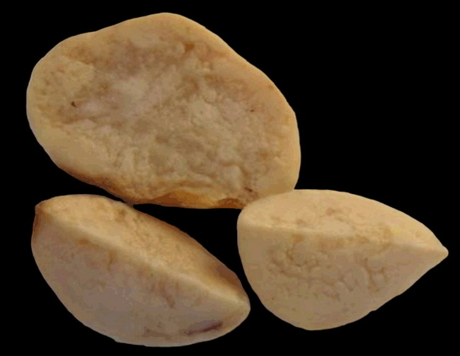
Gross Pathology: Chondroid lipoma is well-circumscribed, lobulated and encapsulated. The size ranges from 1 to 11 cm (median 4 cm). The cut surface is gelatinous, yellow-tan, white, or gray. The location can be in the subcutis or deep soft tissues. Some tumors are located entirely within skeletal muscles.Gross Image: This specimen of chondroid lipoma was removed from a 51 y/o female. She presented with a painless, subcutaneous inframammary mass on the right side of the chest. The resected specimen was a well-circumscribed, encapsulated 8 x 6 x 5 cm mass with a grey-tan, nodular, myxoid cut surface. Image source: Shawarby M.A. et al. Chondroid lipoma, a rare tumor: Report of 2 cases. International Journal of Case Reports and Images 2013;4(10):527-531. Image background changed and image used under Creative Commons attribution 3.0 license.