
Nodular Fasciitis : Intramuscular


Comments:
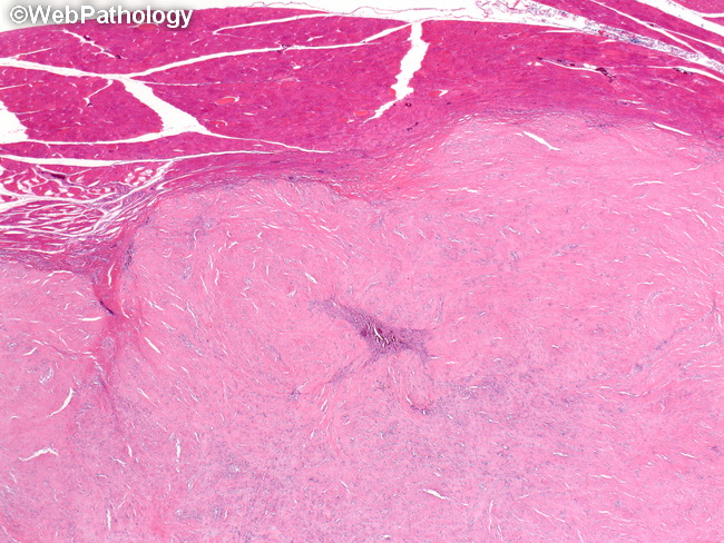
Low-power view showing the interface between the lesion and skeletal muscle bundles in a case of intramuscular nodular fasciitis. Case History: The patient was a 25 y/o female with history of a rapidly growing mass in her deltoid region. The mass had grown to its present size of about 5 cm in three months. It was excised along with a margin of uninvolved muscle. Grossly, the mass was circumscribed and contained entirely within the muscle. It was composed of a bland proliferation of spindle cells arranged in vague storiform pattern. The background stroma was an admixture of loose fibromyxoid areas as well as densely hyalinized areas. The lesional cells were positive for smooth muscle actin and negative for desmin, S-100, h-caldesmon, CD34, and EMA.