
Carcinoid : Gross


Comments:
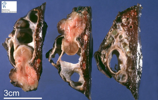
Introduction: Carcinoid tumors make up <1% of all lung cancers (WHO fascicle) and <5% of all primary pulmonary neoplasms (Rosai). They are subdivided into typical carcinoids (TC) and atypical carcinoids (AC) with 70-90% of tumors belonging to the former category. The patients are usually younger than 60 yrs. and they are more common in females and Whites. Carcinoid tumor is the most common primary lung neoplasm in the pediatric age group. TC shows no association with smoking, whereas AC is slightly more common in smokers. MEN1 gene is a risk factor for carcinoids. Diffuse idiopathic pulmonary neuroendocrine cell hyperplasia is considered to be the precursor lesion. Presentation: About two-thirds of lung carcinoids are located centrally near major airways such as main stem or lobar bronchi (as shown in this image). The remaining one-third are peripheral, not associated with a bronchus, and may be asymptomatic. Peripheral tumors are more likely to be atypical carcinoids and often show spindle cell pattern. Centrally located tumors may cause obstructive pneumonia, abscesses, and bronchiectasis. Rare cases present with paraneoplastic manifestations such as Cushing syndrome, acromegaly, and Carcinoid syndrome. Gross: Carcinoids are usually well-circumscribed, round to oval masses filling up the bronchial lumen. They may be sessile or pedunculated. An endobronchial component is seen in about half of typical carcinoids and one-third of atypical carcinoids. The overlying bronchial mucosa is usually intact. They may infiltrate the bronchial wall and extend into the surrounding lung parenchyma. The cut-surface is yellow-gray and may show fibrous septa.